OCT image tips
18/04/2024
Tips
- Quality
-
20 is enough
-
25 is good 😀
-
- 对于pRNFL
- Try to focus better
- In segmentation
- Check the image in the eye & modify if really needed
- Block a part so that it will not be changed by the other part that you are modifying
- QMAC 的 sections 1st 和 25th should be excluded, so modifications in these two sections are not needed
- 做 pRNFL 的时候,可以旋转手柄 joystick 角度多一点,以获得不同角度的 image
Participant
Robert
- White dots in retina
- 双眼都有
- 可能是因为人种原因 melanin 较少
- 可以透过视网膜看到 choroid
Jieying
- In choroid something wrong
- 需转给 ophtalmologist
- Not urgent, but in the next year it would be worse, 早发现早治疗
- Optic disc 不是圆的,这是 astigmatism 的标志
Tiger
- Should go to the doctor
Naroa
- To repeat pRNFL
20240111
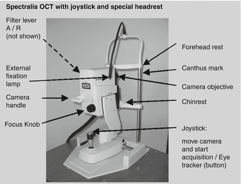
-
how to operate OCT machine
- software: Heidelberg Eye Explorer
- we played as patients in turn and scanned each other’s eye to read the images with Sara’s instructions
-
We have witnessed two participants (control group for Sara’s study) being scanned. The scans involve:
- a) a retina scanning with parameter of QMAC, 25 sections per image
- b) a nerve scanning with parameter of RNFL
- c) an OCTA (OCT-Angiography) scan of vessels, with 512 sections per image
Sara would first check the “display” window of retina scanning to have an overview of the retinal layers, if any line indicator of one layer is out of the actual layer that we can see from our eye, Sara would modify the layer manually to make the image more close to the real retinal layers, esp. the important layers to their study, i.e., the RNFL (retinal nerve fiber layer) and GCL (ganglion cell layer).
After then, Sara would check the nerve image, to check the volume and thickness of the critical layers (the RNFL and GCL).
-
During our stay there, we met Iñigo in person. He explained to us more about the eye anatomy and how to get a high-quality image with the machine. He also pointed out that the machine we were exposed to today cannot analyse data (it can only show us the image qualitatively). If we want to analyze data, we need to use matlab toolbox or the algorithm developed by Iñigo’s group.
take-home notes
- image quality (20~40) over 25 is acceptable, the ones lower than 20 should be excluded
- more practice on the machine operation; to me, it’s not that easy to control the joystick to center the pupil from the beginning
- images can be collected without being contaminated when the participant’s eye leave the machine temporarily (if he/she leaves, the scanning will stop); however, if the retina is still in the viewing window, but part of it is moving, the machine may not catch everything that had happened, so the images would show that some retinal layers are going down on the left/right margin.
20240119
Today Luo and I practiced several times and we operated better than our last time.
We have witnessed Sara scanning 4 participants.
- One of them is a Parkinson’s patient whose retina is quite different from healthy ones in the case of the transparency degree (i.e., we can even see the choroid on the OCT image)
- The other one participant’s scannings could be categorized as control group images even if some age-related degeneration manifestations (high reflectivity spots) were shown.
- One participant is color-blinded since the participant‘s performance on the color numbers test. The nerve scanning showed a weird structure of the retinal vessels in the case of winding out and backwards.
- At the end of today’s apprenticeship, Sara showed us the new software for imaging collection, HEYEX, which is a little different from the Heidelberg Eye Explorer.
Kepa — Sara also pointed out the parameters for scanning right now is customized for their study, including ART frames number, section number, quality, angle, etc. So in our study, we need to define the parameters to suit our investigation objectives, which needs more literature reviews, at least for me.
Iñigo — Attached are the images I took from today (Luo’s retina). Maybe Iñigo can give me some suggestions on the image quality and how to get a better image. Looking forward to your feedback :)
20240125
被试1
没用oct 做的是眼动的cognitive tests
- Hiru 眼动仪 便携式
9 tests 第一个和最后一个是描述图片
中间有 motion emotion memory(1-4objects) tests
有个鹰喙的图片跟Sandra 的实验刺激一样,数据库是啥?
- dive眼动仪
形状test
-
相机 Neur optic是啥
-
color ordering
被试2
Parkinson’s young patient
OD nerve is not equivalent to the one in OS
OCT retina + nerve
OCTA
Eye tracking with dive
Color ordering
(When the eyelash interrupt the image, you should ask the participants to open the eye)
Iñigo will write to Kepa
Protocol of OCT-image resolution
Protocol of MRI
Additional test-behavioral cognitive tests